Measuring Bone Healing
A double-blind study of a new bone-regeneration substrate — and the GPU-accelerated image analysis I wrote to measure how well it worked.
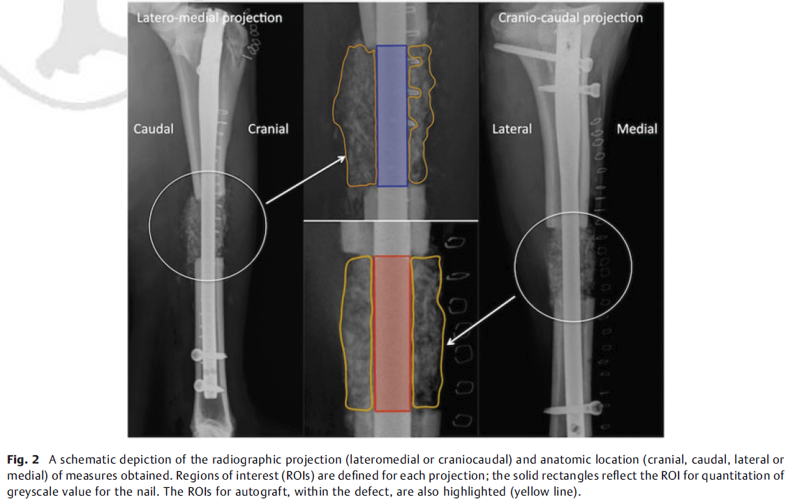
New biomaterials need to be tested and validated before anyone trusts them in a patient. This was a double-blind study of a novel substrate designed to improve bone regeneration and healing, run in a sheep tibial segmental defect model. My part was the measurement: fast, GPU-accelerated radiological image analysis that turns a stack of plain radiographs into numbers a statistician can work with.
I wrote the analysis software in C++, with CUDA for the image processing and OpenGL for the viewer. It extracted optical density, bone density, bone volume and bone mass from the radiographs. The clinical team supplied the qualitative measures — percentage bridging callus, the modified radiographic union score for tibia (mRUST), and locomotory function — and the study compared the two families of measurement against each other.
The headline result was that the qualitative and quantitative measures broadly agreed. Percentage bridging callus and mRUST were almost the same measurement (r = 0.999), while locomotory function correlated poorly with both (r = 0.029 and r = 0.046). Optical density tracked bone density (r = 0.824), bone volume (r = 0.957) and bone mass (r = 0.959) closely enough to be worth collecting: it is cheap to obtain and it contributed real information.
The utilisation of both qualitative and quantitative data, in assessment of the progression of fracture healing, has provided valuable insight. Measures of optical density have been shown to make a substantial contribution to this assessment.
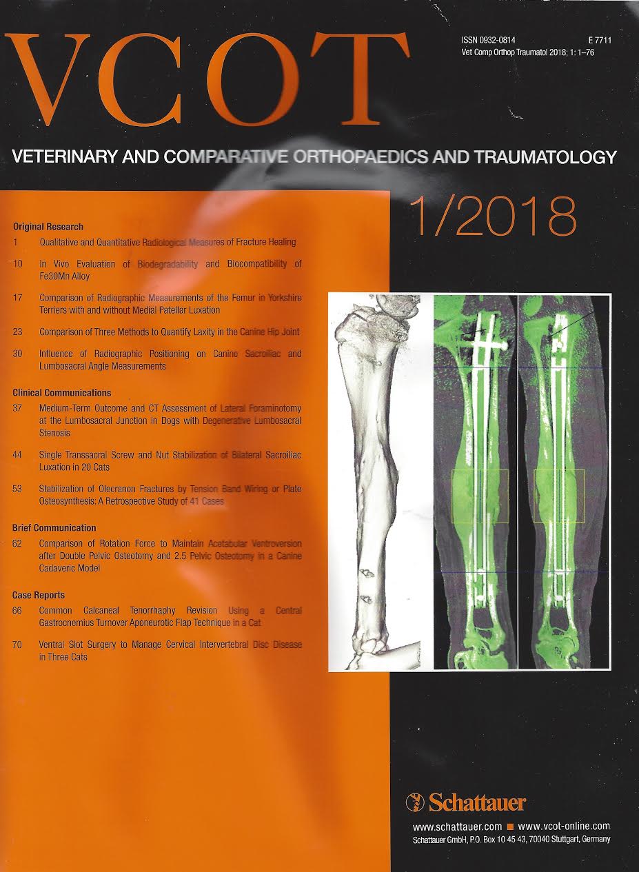
The study was published as Qualitative & Quantitative Radiological Measures of Fracture Healing by J.R. Field and G.S. Ruthenbeck, and it made the cover of the January 2018 issue of Veterinary and Comparative Orthopaedics and Traumatology.